Prenatal nutritional deformities and disease types
A RELATIONSHIP between physical types and certain disease susceptibilities has been recognized by diagnosticians for centuries. The skill of many physicians in reading intuitively and from external signs the nature of their patients' troubles when these could not be classified with precision played an important part in the successful warfare against disease in the period preceding the advance in modern laboratory technique. For many of the old-time physicians, these constitutional qualities were expressed as diathesis. An individual would be recognized as having a phthisical diathesis (a susceptibility to tuberculosis). Similarly, the arthritis group had a rheumatic diathesis. While modern science has undertaken to express its findings numerically, the problem of reducing the diatheses to mathematical formulas has required so many overlappings, that it has been impossible to establish definite limiting boundaries.
In my investigations regarding the types of individuals who develop rheumatic group lesions as a result of dental focal infections, (1) I found that individuals could be divided into very definite groups in which 15.05 per cent with severe lesions belonged to families in which similar disease symptoms had occurred. Evidence was disclosed of a systemic factor that played a controlling role in determining whether or not the individual would be seriously injured from dental focal infections. It became very clear that the soil was quite as important a determining factor as was the type of infection. This finding led me to broaden the scope of my investigations to include a search for control cases that were free from the degenerative processes. I was not able to find these controls in the clinical material afforded by our modern civilization, and therefore extended the search to isolated primitive racial stocks.
Associated with a fine physical condition the isolated primitive groups have a high level of immunity to many of our modern degenerative processes, including tuberculosis, arthritis, heart disease, and affections of the internal organs. When, however, these individuals have lost this high level of physical excellence a definite lowering in their resistance to the modern degenerative processes has taken place. To illustrate, the narrowing of the facial and dental arch forms of the children of the modernized parents, after they had adopted the white man's food, was accompanied by an increase in susceptibility to pulmonary tuberculosis.
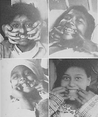
In Fig. 113 will be seen four young people, examined in the tuberculosis wards of the Juneau (Alaska) Hospital for Indians and Eskimos. All exhibited marked evidence of prenatal injury. Note the cuspids erupting outside the line of the arch. The teeth of the upper arch of the boy at the upper left, pass inside the teeth of the lower arch. His upper arch is so narrow that even a finger could not be passed between the lateral walls. These pictures had to be taken with short exposures in the poor light of the wards. They reveal, however, the conditions.
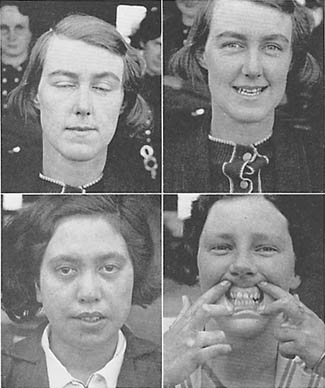
In Figs. 114 and 115 are shown several individuals photographed in the tuberculosis hospital in New Zealand. Note the lack of development of the middle third of the face and the narrowing and lengthening of the face. In several individuals the teeth of the upper arch closed inside the teeth of the lower arch, instead of outside, as in normal persons. Here again, 100 per cent of the young people with tuberculosis gave evidence of injury in the formative period, and 91.2 per cent of the total number of patients were found to have disturbed dental arches.

In Fig. 116 are shown four typical individuals in the tuberculosis hospitals in Hawaii; one in Hilo and the other in Honolulu. In each of these hospitals 100 per cent of the individuals had abnormal development of the face and dental arches.

While we know many of the factors contributing to the nature of diatheses, I have found no data dealing with the forces which determine diatheses, except the influence of heredity. The data I am presenting in this volume, deal with forces other than heredity.
An outstanding advance in organizing the data which relate the physical characteristics of individuals to their disease susceptibilities has been made by the Constitutional Clinic of Columbia University and the Presbyterian Hospital of New York, under the able direction of Dr. George Draper. He has found it necessary in order to study man as a whole, to view him from four different angles: "his form, his function, his immunity mechanism and his psychology." These four attributes he has designated as the "four panels of personality." Dr. Draper has published several communications including two textbooks, one entitled, "Human Constitution," (2) and the other, "Disease and the Man." (3) Dr. Draper has approached this problem from the data provided in the medical clinics, and therefore, from the characteristics of affected individuals, whereas my approach has been through a study of the primitive groups and the physical changes and disease susceptibilities which occur as a result of their modernization. The similarity of our conclusions greatly emphasizes the importance of the findings of each. Dr. Draper has emphasized the importance of the face and of the dental arches in the general matter of susceptibility to disease. He closes one of his chapters, entitled The Relation of Face, Jaws and Teeth to Human Constitution and Its Bearing on Disease, as follows:
The lessons which we have learned from these observations, however, is that the face and jaws hold much information of value to the student of the human being. As clinicians in the field of internal medicine we have been taught to observe the gums and teeth in order to detect possible foci of infection. But for the student of clinical organismalism, the teeth and jaws hold much valuable information about the total personality. For the worker in the dental branch of medicine it would seem that an unusual opportunity is offered for extending such observations and correlations. It may very well be that the dental student who becomes interested in the relation of the mouth to the organism may form a most important link with the responsibilities of internal medicine.
The more we come to view man as a totality, as an organism which functions as a whole and not as a collection of separate elements, the more do all the special branches of medicine become fused with the general concept which forms the basis of this discussion, namely, the relation of the human organism as a whole to those various reactions of maladjustment with environment which we call disease.
It is clear that a definite association of abnormal facial patterns with specific disease susceptibilities exists. From my studies it is also clear that these abnormal facial patterns are associated with influences resulting from a change in the nutrition of the parents of the individual. We are at this point concerned with the forces that underlie these phenomena.
In approaching this problem as it applies to human beings, much can be learned from a study of domestic and wild animals. Until recent years it has been common knowledge among the superintendents of large zoos of America and Europe that members of the cat family did not reproduce efficiently in captivity, unless the mothers had been born in the jungle. Formerly, this made it necessary to replenish lions, tigers, leopards and other felines from wild stock as fast as the cages were emptied by death or as rapidly as new stock should be added by enlargement.
The story is told of a trip to Africa made by a wild animal specialist from the London zoo for the purpose of obtaining additional lions and studying this problem. While in the lion country, he observed the lion kill a zebra. The lion proceeded then to tear open the abdomen of the zebra and eat the entrails at the right flank. This took him directly to the liver. After spending some time selecting different internal organs, the lion backed away and turned and pawed dirt over the carcass which he abandoned to the jackals. The scientist hurried to the carcass and drove away the jackals to study the dead zebra to note what tissues had been taken. This gave him the clue which when put into practice has entirely changed the history of the reproduction of the cat family in captivity. The addition of the organs to the foods of the captive animals born in the jungle supplied them with foods needed to make reproduction possible. Their young, too, could reproduce efficiently. As I studied this matter with the director of a large lion colony, he listed in detail the organs and tissues that were particularly selected by animals in the wilds and also those that were provided for animals reproducing in captivity. He explained that, whereas the price of lions used to be fifteen hundred dollars for a good specimen, they were now so plentiful that they would scarcely bring fifteen cents. If we observe the parts of an animal that a cat eats when it kills a small rodent or bird, we see that it does not select exclusively the muscle meat.
During my biological investigations using animals, I have had barn rats gnaw their way into the room where the rabbits were kept and kill several animals during a night. On two different occasions, only the eyes of the rabbits had been eaten, and the blood may have been sucked. On another occasion the brains had been eaten. It was evident that these rats had a conscious need for special food elements that were provided by these tissues.
No phases of the problem of physical degeneration can be as important as knowledge of the forces that are at work and the methods by which they operate. It is clear from the data presented in previous chapters that these forces can become operative with sufficient speed to make a difference in two generations, one succeeding the other. It is also clear from preceding data that these forces originate in the change of the nutrition of the parents.
A chemical analysis of the food (Chapter 15), discloses a marked reduction in the intake of some of the vitamins and minerals in individuals who are undergoing a degenerative process.
Many investigators have presented important data dealing with the role of vitamin A in prenatal as well as postnatal growth processes. It is known that the eye is one of the early tissues to develop injury from the absence of vitamin A, hence the original name for this vitamin was the xerophthalmic vitamin. The importance of vitamin A to the eye, and the fact that this vitamin is stored in eye tissue have been emphasized by several investigations.
Wald (4) in discussing vitamin A in eye tissues states:
Extracts of eye tissue (retina, pigment epithelium and choroid) showed the characteristic vitamin A absorption band at 620 mu. with the SbC13 test and were also potent in curing vitamin A deficient rats. The concentration of vitamin A was very constant for different mammals, at about 20 Y per g. dry tissue. Values for frog tissues were much higher.
This comment is of interest in connection with the observation that I have previously quoted regarding the raid of barn rats on the rabbit cages in the stress of deep winter. While it has been shown that vitamin A is essential for the normal function of eyes, its role in the formation of eye tissues has not been clearly understood. Probably the most sensitive procedure for testing the depletion of vitamin A in domestic animals today is by observing their behavior in semi-darkness.
Edward Mellanby (5) has presented important new data dealing with vitamin A deficiency and deafness. He states in an abstract of a paper read before the Biochemical Society, in London in November 1937, the following:
In previous publications I have shown that a prominent lesion caused by vitamin A deficiency in young animals, especially when accompanied by a high cereal intake, is degeneration of the central and peripheral nervous systems. In the peripheral system it is the afferent nerves which are principally affected, including the eighth nerve, both cochlear and vestibular divisions. It has now been possible to show that vitamin A deficiency produces in young dogs degenerative changes in the ganglia, nerves and organs of both hearing and balance inside the temporal bone. All degrees of degeneration have been produced, from slight degeneration to complete disappearance of the hearing nerve. The nerves and ganglion cells supplying the organ of Corti are more easily damaged than those of the vestibular division. As might be expected when once the spiral ganglion cells have disappeared, additions of vitamin A to the diet have no effect, and the organ of Corti remains completely denervated. The "inattention" of dogs on these diets which I previously ascribed to cerebral defect is undoubtedly due to deafness. It now remains to determine whether these results may be extended to explain certain forms of deafness in man.
The serious effects of deficiency in vitamin A on pregnant rats has been investigated and reported by Mason (6) as follows:
Abnormalities are described in the pregnancies of rats maintained on diets deficient in vitamin A in varying degree. Prolongation of the gestation period up to 26 days in severe cases and a long and difficult labour which might last 2 days and often resulted in death of both mother and young were characteristic.
Defects due to deficiencies in vitamin A in the diet of dairy animals, (pregnant cows, their offspring and normal calves fed on the milk from those cows) have been reported upon by Meigs and Converse (7) as follows:
In 1932 we reported from Beltsville evidence that farm rations frequently fed to calves may be dangerously low in vitamin A, and that milk produced by cows fed hay which has lost its green color may be an unsafe source of vitamin A in the calf ration. This preliminary paper reported results dealing with four cows which had been fed over two years on a good grain mixture and late-cut low-color timothy hay. Of six calves born of these cows, two were dead, one was unable to stand and died shortly after birth, and three were both weak and blind. The fact that cows so fed were unable to properly nourish their calves before birth led to the question whether the milk from these cows might not be deficient for normal growth of calves from other cows fed on rations adequate in vitamin A. This preliminary report included results on three normal calves fed milk from those cows fed low-color timothy hay. The three calves died at fifty-seven, sixty-two, and seventy-one days of age, respectively.
Since growth, prenatal and postnatal, is related directly to the pituitary body, we are specially interested in the knowledge available on the functioning of this gland.
The work of Barrie (8) throws important light on this subject. He has reported as follows:
Partial deficiency of vitamin E as shown in the female rat fed on a diet containing only a trace of vitamin E but which is otherwise complete, results in the prolongation of gestation, which may be continued as long as 10 days beyond the normal period. The offspring under these conditions are abnormal. These young may develop slowly and be thin and undersized in spite of sufficiently profuse lactation, or they may become extremely fat and develop a leg weakness and carpopedal spasm about 18 days after birth. Animals of both types have thin skulls and short silky fur. Complete E deficiency in the adult also produces the soft fur and imperfectly calcified skull. Partially E deficient animals occasionally give birth to a litter but fail to lactate.
The changes observed are similar in several ways to those produced by hypophysectomy (surgical removal of pituitary gland). Marked degranulation of the anterior pituitary is found in both the abnormal young and the adult sterile animals. Lack of vitamin E therefore produces a virtual nutritional hypophysectomy in the young rat.
The inability of various species to complete gestation when vitamin E is not provided in adequate quantity has been reported by many investigators. A. L. Bacharach, E. Allehorne and H. E. Glynn have used their study of this factor as a means of estimating the quantity of vitamin E in the diet. (9) They state:
A diet suitable for work on vitamin E causes female rats, fed on this diet from weaning, to show a gestation-resorption rate not significantly differing from 100 per cent; when the diet is supplemented with adequate amounts of vitamin E, also administered from weaning, the live litter rate does not differ significantly from 100 per cent. It is found that, on such a diet, the percentage of implantations (conceptions) is markedly different between animals that have been submitted to a single gestation-resorption, owing to lack of vitamin E, and animals that are mated for the first time. The authors suggest that the process of undergoing gestation-resorption brings about some deep-seated changes in the reproductive mechanism of the rat, of a kind not hitherto recognized as characterising the vitamin E deficiency syndrome.
One of the outstanding changes which I have found takes place in the primitive races at their point of contact with our modern civilization is a decrease in the ease and efficiency of the birth process. When I visited the Six Nation Reservation at Brantford, Ontario, I was told by the physician in charge that a change of this kind had occurred during the period of his administration, which had covered twenty-eight years and that the hospital was now used largely to care for young Indian women during abnormal childbirth (Chapter 6).
A similar impressive comment was made to me by Dr. Romig, the superintendent of the government hospital for Eskimos and Indians at Anchorage, Alaska. He stated that in his thirty-six years among the Eskimos, he had never been able to arrive in time to see a normal birth by a primitive Eskimo woman. But conditions have changed materially with the new generation of Eskimo girls, born after their parents began to use foods of modern civilization. Many of them are carried to his hospital after they had been in labour for several days. One Eskimo woman who had married twice, her last husband being a white man, reported to Dr. Romig and myself that she had given birth to twentysix children and that several of them had been born during the night and that she had not bothered to waken her husband, but had introduced him to the new baby in the morning.
Sherman, (10) who has made many important contributions to our knowledge of vitamin A, has shown in a recent communication that an amount of vitamin A sufficient to support normal growth and maintain every appearance of good health in animals, may still be insufficient to meet the added nutritive demands of successful reproduction and lactation. With the failure to reproduce successfully, there usually appears in early adult life an increased susceptibility to infection, and particularly a tendency to lung disease at an age corresponding to that at which pulmonary tuberculosis so often develops in young men and women. He states, further, that vitamin A must be supplied in liberal proportions not only during the growth period but during the adult period as well, if a good condition of nutrition and a high degree of health and vigor are to be maintained.
Hughes, Aubel and Lienhardt (11) have shown that a lack of vitamin A in the diets of pigs has resulted in extreme incoordination and spasms. They also emphasize that gilts bred prior to the onset of the nervous symptoms either aborted or farrowed dead pigs.
Hart and Gilbert (12) have shown that the symptoms most commonly seen in cattle having a vitamin A deficiency are the birth of dead or weak calves, with or without eye lesions. They report also a condition of newborn calves which simulates white scours, and the development of eye lesions in immature animals.
Hughes (13 has shown that swine did not reproduce when fed barley and salt, but did so when cod liver oil was added to this food.
Sure (14) has shown that a lack of vitamin A produces in females a disturbance in oestrus and ovulation, resulting in sterility. Further, he states, that resorption of the fetus may be produced by lack of vitamin A, even on a diet containing an abundance of vitamin E, which is known as the anti-sterility vitamin.
One of the most important contributions in this field has been made by Professor Fred Hale, of the Texas Agricultural Experiment Station, at College Station, Texas. He has shown that many physical deformities are readily produced by curtailing the amount of vitamin A in the ration of pigs. He produced fifty-nine pigs (15) that were born blind, every pig in each of six litters-where the mothers were deprived of vitamin A for several months before mating and for thirty days thereafter. In pigs, the eyeballs are formed in the first thirty days. He found, as have several others, that depriving pigs of vitamin A for a sufficient period produced severe nerve involvements including paralysis and spasms, so that the animals could not rise to their feet. He reported that one of these vitamin A deficient pigs that had previously farrowed a litter of ten pigs, all born without eyeballs, was given a single dose of cod liver oil two weeks before mating. She farrowed fourteen pigs which showed various combinations of eye defects, some had no eyes, some had one eye, and some had one large eye and one small eye, but all were blind.
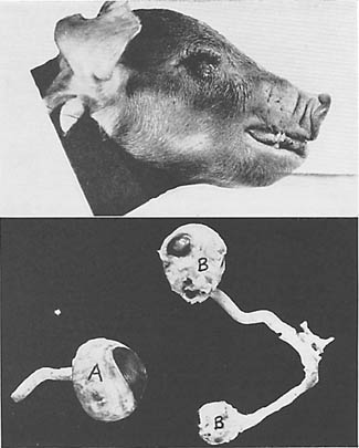
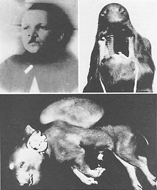
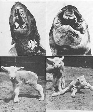
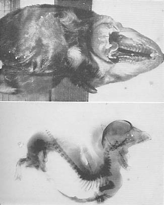
In Fig. 117 I am able through Professor Hale's kindness, to show an eyeless pig and a normal eye of a pig (at the left) and (at the right) a pair of incomplete eyes from a pig born in the litter just mentioned. This one dose of vitamin A made possible the partial formation of optic nerves and eyeballs. A typical eyeless pig is shown in Fig. 118 (lower). Note its deformed ears. Among the many physical injuries which develop in the pigs born to sows fed on a diet deficient in vitamin A are serious defects of the snout, dental arches, eyes and feet. This pig was born without eyeballs. It also had club feet and two tumors. In Fig. 118 (upper right) is shown a pig with cleft palate, and in Fig. 119 (upper right) one with double hairlip. In Fig. 118 (upper left) is shown a boy with cleft palate and defective eyes. One of the very important results of Professor Hale's investigations has been the production of pigs with normal eyes, born to parents both of whom had no eyeballs due to lack of vitamin A in their mother's diet. The problem clearly was not heredity. Two litters, one containing nine pigs and the other eight, which were born to mothers that had been deprived of vitamin A before mating and for thirty days thereafter, produced the following lesions: All had complete absence of eyeballs; some lacked development of the opening of the external ear; others had cleft palate, harelip, displaced kidneys, displaced ovaries, or displaced testes.
It is of interest that in October, 1935, Professor Hale reports that the Texas Agricultural Experiment Station was informed that a litter of fourteen pigs had been born blind in June, 1935, on a farm at Ralls, Texas. Of these, six pigs were raised and brought to the Station for further study. The farmer owning the pigs stated that no green feed was available on his farm from March, 1934, until May, 1935. It will be noted that this condition paralleled the experimental conditions at the station under which by restricting vitamin A before and immediately after gestation fifty-nine pigs were produced without eyeballs.
Professor Hale reports that in April, 1935, a litter of seven pigs were born blind at McGlean, Texas, which was suffering from drought conditions, similar to those at Ralls. The litter and dam were purchased by the Experimental Station. Matings were made between blind pigs. These were fed rations containing ample vitamin A, and normal pigs with normal eyeballs were produced. Even the mating of a blind son with his mother who had produced him when on deficient diet, produced only normal pigs when both had ample vitamin A. He states, "If an hereditary factor had been the cause of this congenital blindness, these matings would have produced some blind pigs, even if vitamin A were present in the ration." The problem of congenital cleft palate has been very embarrassing to those parents whose children have been so afflicted. It is of interest that breeders of fancy dogs are frequently embarrassed by having this problem develop in their kennels or among litters born to parent stock obtained from their kennels.
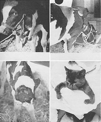
In Fig. 119 (upper left) I have shown a water spaniel pup with cleft palate. This pup was not able to nurse because of the impossibility of producing suction without a palate. When fed artificially the milk was expelled through the nostrils. The mother had given birth to two previous litters all of which were dead at birth or died soon after. Her diet had been reinforced with mineral calcium phosphate in tablet form hoping to insure normal offspring. This is not nature's method.
I am informed by a veterinary that he has experienced trouble with harelip, cleft palate or serious facial deformities among dogs that are pets in homes where they are lavishly coddled and fed the things they like best. He stated that he has more trouble with head defects in bulldog pups than any other breed.
There are few if any problems connected with modern degeneration on which so much light is thrown as that supplied by recent investigations on the problems of paternal responsibility for defects in the offspring. There are several reasons for this. Because the mother has the sole responsibility for the nourishment of the fetus during the formative period and she alone provides the handicaps incident to the process of birth, it is very natural that defects are practically all interpreted as being associated with these processes. This unfortunately, has been embarrassed further by the fact that since distortions in behavior do not appear until sometime after birth, normality was largely assumed to be present up to the time of their appearance, and therefore of necessity would be contributions from the child's environment. As such they would be subject naturally to treatment by applying influences to change the mental environment. Hence the entire problem of the role of the sex cells through controlling the architecture of the body including the brain has been largely overlooked. Very important light is thrown on this problem in the data provided on the pup shown in Fig. 120. This shows a dachshund pup with cleft palate and a very severe spinal deformity. This is not unlike deformities frequently seen, or unlike that of the pup shown in Fig. 119. The highly significant circumstance is the fact that these same deformities not only appeared also in another pup of this same litter but in one pup in each of three other litters about the same time. While four mothers were involved these four litters were all sired by one father. The paternal responsibility is clearly established.
At the right in Fig. 119 a lamb is shown with club feet and two lambs without eyeballs. I am told that in some of the sheep raising states quite a number of deformities occur in lambs at birth. One writer states:
These deformities may maintain themselves in any number of ways. We have them two-headed, 5 and 6 legs, 2 tails, born without eyes, hermaphrodites, born with ribs on one side and any number of such deformities. A common deformity in sheep is under-shot and over-shot jaws. This simply means that the upper jaw extends farther to the front that the under jaw and the front teeth do not meet. This is called over-shot and the reverse is called under-shot.
It will be noted that a common deformity in sheep relates to an under-development of either the upper or lower jaw which is one of the most common expressions of disturbed development in humans.
Some illustrations of the deformities that may occur in domestic animals are shown in Fig. 121. Above are seen two cows each with an extra foreleg attached to the shoulder; below, to the left is shown a double-faced calf which also had a cleft palate; to the right a cat with deformed legs.
In corresponding with Professor Hale, I inquired whether they had information as to the effect of vitamin A deficiency on the sire. He replied, "If we reduceed the vitamin A content of the body of the sire, he would become sterile and, therefore, we could not try this procedure." The question arises as to what the effect would be of a less severe depletion of vitamin A of both parents.
It is a very easy matter to place the full responsibility on the mother, when defects develop in the children. These data indicate that either parent may contribute directly to certain of the defects of the children, due to defects in the germ plasm.
A practical case from my field studies includes a full-blooded Eskimo woman who was married twice, the second time to a white man, by whom she had several children. She had insisted on selecting and preparing the native foods for herself, though she prepared the white man's imported foods for him. With a total of twenty-six pregnancies she did not have any tooth decay. He had rampant tooth decay, and a marked abnormality in the development of face and of the dental arches. Several of the children had incomplete development of the face and of the dental arches. One of the girls who was married had very narrow dental arches and nostrils and a typical boyish type of body build. Unlike her mother, this girl had a very severe experience in the birth of her only child and insisted that she would not take the risk of having another. Several daughters have narrow arches. The question arises whether the deficient nutrition of the father may have been the contributing factor in the injury of their children.
New light is being thrown on human problems by animal investigations. One study that is particularly instructive has been conducted by McKenzie and Berliner of the University of Missouri. (Bulletin No. 265.) The experimental animals used were sheep. Their studies indicated to them that they could predict the level of fertility of the sire by studies made on the seminal fluid. They found that the percentage of abnormal spermatozoa increased to 84 per cent under unfavorable conditions and reduced to less than 15 per cent under favorable conditions. When the unfavorable conditions characterized by these abnormal spermatozoa prevailed the females did not conceive. High temperature proved to be an important controlling factor as indicated by variation in percentage of abnormal sperm. In one breed the average variation was from 2.5 per cent in January to 22 per cent defective in August; in another breed from 18 per cent in January to 73 per cent defective in August. They also found that the same result could be produced in the winter by keeping the sires in heated rooms or by applying heat externally. They further found that when the percentage of abnormal sperm was high a condition of sterility was apparently established. In a personal communication from Professor McKenzie he states that their inference is that with a high percentage of abnormal sperm the ova either are not fertilized or that fertilization takes place and that prenatal death occurs in a certain number of the cases, and that there is an appreciable prenatal death loss in sheep, horses, cattle, and swine. I have referred to the data presented by Mall indicating that 15 per cent of human conceptions in England are expelled as deformities in the second and third month of gestation. McKenzie also presents data relating to swine in which a sire that showed a high percentage of abnormal sperm was bred to two groups of sows of similar breed. One was maintained in a barn without access to pasture, the other received similar rations plus access to pasture. The group without pasture produced a high percentage of mummies, small litters, and abnormal pigs (open abdominal walls with viscera protruding and exposed spine in the region of the lumbar vertebrae). The second group on the same food with the addition of pasturage produced normally large-size litters without mummies. The following year this experiment was repeated reversing the groups of sows and using a similar high percentage of defective sperm in the same sire. The same results occurred, with the group maintained without access to pasture. This emphasizes the need for the vitamins and minerals that are provided in natural foods, particularly vitamin A and seems to relate to the studies of Hale in which the absence of vitamin A produced gross defects. These studies on domestic animals strongly emphasize the necessity that both parents shall have adequate nutrition before conception occurs and subsequently for the mother.
Until very recently there has been exceedingly little literature dealing with the factors contributing to the abnormal development of fetuses in either domestic animals or humans. Williams, who is one of the few students of this problem as it relates to domestic animals, in referring to this phase has commented that there is no treatise in the English language on teratology (development of monsters) in domestic animals. (16) He emphasizes that these defects are growing in numbers and economic importance. He refers to Burki's studies in Switzerland indicating that this difficulty has constantly and enormously increased in his territory. This veterinary has asked "Are our cows degenerating?" It is of interest that a type of deformity commonly recognized among veterinaries as a distortion of the head presenting a lack of development of the bones of the middle part of the face including the upper jaw is commonly called "bull-dog calf." This is particularly important in connection with the frequent human deficiency of lack of development in the middle third of the face. Williams emphasizes the common knowledge that Boston bull-dogs are not prolific and because of the difficulty of giving birth to their young, some veterinarians resort routinely to caesarian section. Fortunately, seriously deformed specimens are seldom born alive and usually do not continue to term. Williams states that Loje has observed ten grossly deformed calves of a particular type that were traced to the same sire; also a series of five with this deformity reported by Hutt were traced to one sire. Among the deformities in domestic animals cleft palate or absence of palate is very common. These studies on domestic animals strongly emphasize two facts; first, that deformities among these animals are very similar to those that develop in humans, and second, that the defects are largely related to the original germ cells and that the male may provide the defect quite as well as the female. Several of the primitive races have understood and provided against these mishaps.
Moench and Holt of the Cornell Medical School and New York Hospital (17) have made important studies on humans and have found a very high incidence of sterility when abnormal forms of spermatozoa reached 25 per cent. Among the abnormal forms they found one particular family with a particular type of abnormal sperm reaching 12 per cent. Their breeding record was decidedly bad and fetal malformations repeatedly occurred. In their group, in 63 sterile matings the men were normal 21 times and abnormal 37 times. They listed over 40 different abnormal or deformed types of sperm. They conclude:
- In a normal semen the abnormal sperm heads do not exceed 19 to 20 per cent.
- When the sperm head abnormalities reach 20 to 23 per cent, impaired fertility can be assumed.
- When the sperm head abnormalities are above 25 per cent, clinical sterility is usually present.
An important contribution to the question of the relation of the germ cells of the father to the type of defects that are prevalent in his family history has been made in recent studies conducted in Germany. (18)
Two German surgeons concerned with enforcing the sterilization law in Berlin have taken advantage of their opportunities to make an important contribution to the study of fecundity.
Several studies have been published previously, of the spermatozoa of normal men. This book offers a study of the spermatozoa of men who had been diagnosed as hereditarily defective. At the time of the operation, the upper part of the vas deferens was washed out, and the sperm thus obtained were studied in great detail, 20 different types of abnormality being distinguished.
In their control studied, the normal man is found to produce 19 per cent of morphologically defective sperm. By contrast, the chronic alcoholic patient produces 75 per cent of defective sperm.
Previously it has been assumed that as much as 25 per cent or 30 per cent of abnormal sperm were a good evidence of lowered fertility if not of sterility. But the fertility of these chronic alcoholics was not diminished, the authors claim.
In cases of inherited mental defect, 62 per cent of abnormal sperm were produced, accompanied by low fecundity,--as is the case generally with the mentally defective male. On the other hand, in hereditary deafness, 62 per cent and in hereditary blindness 75 per cent of defective sperm appeared.
Epilepsy and schizophrenia (dementia praecox) both supposed to be constitutional in origin, show less abnormality of spermatozoa (58 per cent and 54 per cent respectively) than blindness and deafness.
The fact that in the past almost the entire emphasis of change in physical, mental and moral qualities has been assigned to the forces of heredity, strongly emphasizes the lack of information regarding the nature of the forces at work and the points at which their influence constitute the determining factors. The data previously presented in this chapter dealing with the role of vitamins A and E throw valuable new light on this phase. It is important, however, that new data are available dealing with the factors which injure the germ cells and which Tredgold has spoken of as "poisoned germ cells."
In a personal communication received from Professor T. S. Sutton of the College of Agriculture, Ohio State University, he makes the following observation:
For several years we have been interested in the study of the effects of vitamin A deficiency. Right now our main consideration is the effect of this deficiency on reproduction. We find that a diet low in vitamin A will cause reproductive failure, which seems to be caused chiefly by a degeneration of the germinal epithelium of the gonad. This is particularly true in the case of the male. I think we have rather convincing evidence that this is a direct dietary damage to the gonad (ovary or testis), rather than a disruption of the endocrine balance which might result in sterility as appears to be the case in vitamin B deficiency.
The degenerative processes which occur in nerve tissues due to vitamin A deficiency have been studied by Professor Sutton and his associates. (19) They have been able to show detailed progressive degeneration within nerve fibers as a result of vitamin A deficiency.
In the light of the newer information it is quite clear why we may have anyone of the following distinct expressions in the reproductive process, namely, physical excellence of succeeding generations (such as obtains among many of the primitive races as I have shown); complete reproductive failure or sterility or partial failure resulting in defectives of various types at the borderline between these two phases. It is the rapid and progressive increase in this last group which constitutes the progressive degeneration of our modern civilization.
One of the main problems in this study has to do with the relation of nutrition to the modification of the growth of the child, both in its formative period and in the stage of adolescence. I have shown that in many of these primitive racial stocks there occurs in the first generation after the displacement of native foods by imported foods a marked change in facial and dental arch forms. These changes happen most frequently in the later children in the families and come about notwithstanding the impact of heredity through all the previous generations of excellent physical development. Clinically, the evidence is abundant, that this change occurs in these primitive racial stocks regardless of color, geographic location, temperature, and climate. We are apparently dealing here with a factor which, while it may be related to the germ plasm and to the prenatal growth period, clearly involves other forces than those that are at work in the case of hereditary defectives. Since these changes have to do directly with disturbances in growth of the head, particularly of the face and of the dental arches, we are concerned with such evidence as may be available as to the nature of the forces that readily affect the anatomy of the skull.
The general architecture of the body is apparently determined primarily by the health of the two germ cells at the time of their union. This architectural design may not be completely fulfilled due to interference with nutritive processes both before and after birth. In this large problem of the relationship between physical design of the body and resistance or susceptibility to disease, we may have determining factors operating at different periods in prenatal and postnatal growth. The accumulating evidence strongly emphasizes that disease susceptibility is a widely variable factor and associated with certain types of developmental disturbances.
In a discussion of tuberculosis susceptibility before a meeting of specialists in that field, emphasis was placed upon the fact that other factors than the bacteria played controlling roles in the matter of susceptibility to tuberculosis.
Weisman has recently made an important contribution to the problem of physical development and susceptibility to tuberculosis. He has presented (20) statistical data indicating the type of chest deformity which predisposes an individual to tuberculosis. He states:
In a study previously made on the shape of the normal and of the tuberculosis chest, it was found that the average normal chest was flat and wide and that the average tuberculous chest was deep and narrow. It was also shown that the deep chest was an underdeveloped, primitive type of chest, resembling an infant's chest in shape. Later studies on the shape of the chest and on environment showed that children from the poorer socio-economic environments had on the average a deeper chest, weighed less and were shorter than the children from the higher socio-economic levels. An investigation recently made on the incidence of tuberculosis in the various school districts in Minneapolis revealed that there is a very high incidence of tuberculosis among the children from the slums where the deep chest prevails. Ten times as many cases of tuberculosis were reported from a school district which is perhaps the poorest in the city as were reported from the best school districts.
This study, which shows that there is a definite correlation between the deep chest and the positive reaction to tuberculin adds one more link to the chain of evidence supporting the contention that the deep chest is more or less associated with tuberculosis. It also helps to explain why there is such a high incidence of tuberculosis among the poor in the slum districts. The children in the slums are physically underdeveloped. They are not only shorter and lighter but they have on the average a deep, primitive, infantile type of chest, one that has not gone through the normal process of development. Even the new-born and infants are shorter and lighter and have a deeper chest than the average infant from a better environment.
It is important to note that Dr. Weisman associated the type of chest which predisposes the individual to tuberculosis, with a prenatal condition since he states "even the new-born and infants are shorter and lighter and have a deeper chest than an average infant from a better environment." This work throws important light on why in the primitive groups the children born to parents who are living on the imported nutrition lower in vitamins and minerals than the native foods, not only showed a greatly increased incidence of tuberculosis over the children born to parents on the native diet but also proved to be those individuals who, in facial and dental arch form, presented positive evidence of prenatal injury. We also have a direct explanation for the observations that have been emphasized by Dr. George Draper, that physical form has a direct relationship to disease susceptibility of certain types, frequently spoken of as diatheses.
It is important in this connection to call to mind the statement made by some clinicians that tuberculosis patients with narrow nostrils tend to make a poor fight. These narrow nostrils are clearly related to prenatal injury resulting primarily from defective germ cells which determine the architecture, not heredity but intercepted heredity.
It is very easy to understand the effects of gross physical lesions such as the absence of eyeballs, harelip and cleft palate. These defects can readily be seen. The problem is very different, however, when we are dealing with disturbances of function due to minute anatomical lesions in either external or internal organs such as the brain. These latter will be discussed in the next chapter.
References
Next
Table of Contents
Back to the Small Farms Library